
In this episode, the body is invaded by two pathogens from contaminated raw seafood: Vibrio bacteria and the Anisakis parasite. Eosinophil tries unsuccessfully to kill the Vibrio bacteria as it invades the intestine. After the neutrophils kill the Vibrio bacteria, the other cells mock Eosinophil for being a weakling. Soon after; though, the Anisakis worm invades the stomach. Despite pleas from other cells to run away, Eosinophil faces the parasite alone and kills it in one stab. Much to Eosinophil’s embarrassment, the other cells commend her for killing the parasite and apologise for underestimating her.
How do neutrophils and eosinophils compare in killing bacteria and parasites respectively? Join us as we look at the killing mechanisms of these two distinct white blood cells.
Vibriosis: a bacterial infection
| Schematic | Microscopic | Anime |
 |  |  |
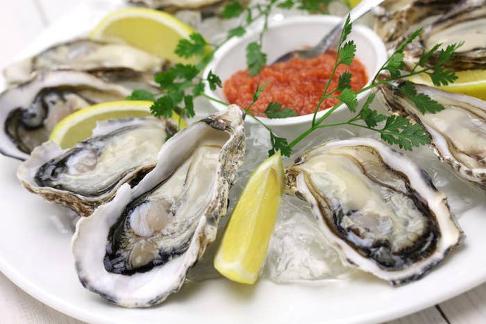
Vibriosis is an infectious disease caused by pathogenic Vibrio bacteria, typically Vibrio parahaemolyticus which invaded the intestine in the anime episode. Vibrio vulnificus is another bacteria that causes vibriosis, typically causing skin infections and flesh-eating disease around open wounds.These bacteria are found in estuaries (where salt water meets fresh water) and coastal waters, particularly the summer months when water temperatures are warmer. Here, molluscs such as oysters catch bacteria on filters as they pump water through gills to collect oxygen and food, concentrating the bacteria up to 100-fold. When consumed raw or undercooked, these molluscs cause 90% of vibriosis cases. Within 24 hours, the person suffers from diarrhoea with abdominal cramping, nausea and vomiting. However, the infection is non-lethal, usually subsiding after three days. In the meantime, fluids lost from diarrhoea can be replaced by drinking plenty of liquids containing electrolytes (a mixture of salts and sugars).

Did you know? Vibrio cholerae is the bacteria that causes cholera. This disease is far serious as it causes rapid body fluid loss due to diarrhoea and vomiting, leading to dehydration, shock and death. Cholera is prevalent in developing countries with poor sanitation, producing 2.9 million cases and 95,000 deaths per year.
How do neutrophils kill bacteria?

When Vibrio bacteria such as Vibrio parahaemolyticus invade the body, neutrophils migrate from the bloodstream to the infected area. Here, neutrophils engulf and degrade Vibrio bacteria via phagocytosis. Initially, neutrophils detect host proteins that coat bacteria such as antibodies and complement proteins. As these host proteins interact with receptors on neutrophils, they engulf the bacteria.
From here, neutrophils kill bacteria in three ways:
- Production of highly toxic reactive oxygen species (ROS): neutrophil enzymes such as NADPH oxidase and myeloperoxidase (MPO) produce reactive chemicals called ROS such as superoxide anion (O2–), hydrogen peroxide (H2O2) and hypochlorous acid (HOCl). ROS species are highly toxic to bacteria, killing them by inactivating bacterial proteins and impeding DNA replication.
- Fusion of bacteria-containing vacuoles with neutrophil granules: neutrophil granules contain various enzymes such as neutrophil serine proteases (NSPs) and bactericidal/permeability-increasing protein (BPI) as well as antimicrobial proteins such as lactoferrin, lysozyme and calprotectin. These mediators break down bacteria or reduce their virulence and initiate inflammatory responses.
- Neutrophil extracellular traps (NETs): neutrophils can sometimes release a mesh-like structure made of DNA and histones that is studded with granule-derived antimicrobial peptides and enzymes. NETs are designed to trap and degrade bacteria outside the neutrophil.

Anisakiasis: a parasitic infection
| Schematic | Microscopic | Anime |
 |  |  |
Anisakiasis, or herring worm disease, is an infectious disease caused by larvae of the worm Anisakis simplex. These larvae enter the human body by consuming raw or undercooked contaminated seafood in dishes such as sashimi and ceviche where they attach to and invade the gastrointestinal walls of the stomach and intestine.

Anisakis simplex worms have a life cycle that normally does not involve humans. Adult worms within the stomach walls of sea mammals such as whales and dolphins mate and lay eggs which are released in faeces. In the sea bed, larvae develop inside eggs (L1 stage) which eventually hatch (L2 stage). The larvae are ingested by small crustaceans such as krill and plankton where they reach an infective stage (L3) by the time they are consumed by fish and squid. Normally, when fish and squid carriers are consumed by marine mammals, the larvae matures to the L4 stage and grow to adult worms, repeating the life cycle. Humans can be the accidental hosts of these larvae; however, when they consume raw or undercooked contaminated seafood. Here, the larvae cannot fully develop into worms but they can still invade the gastric and intestinal walls to cause illness. Symptoms produced within 12 hours of consuming the larvae include abdominal pain, nausea, vomiting and diarrhoea. These symptoms arise due to the increased motility or movement of the gastrointestinal tract as it tries to expel the larvae from the body.
How do eosinophils eliminate Anisakis larvae?

As soon as Anisakis larvae attach and burrow themselves to the gastrointestinal wall of the stomach or intestine, they release chemicals that attract eosinophils. When they reach the larvae, eosinophils release many enzymes such as major basic protein, eosinophil cationic protein and eosinophil-derived neurotoxin that damage and kill larvae. These enzymes can also damage the surrounding tissue, producing dead tissue around the dying larvae. Helper T cells also release cytokines that promote eosinophil activity. Most notably, IL-5 from helper T cells promote eosinophil proliferation and activation and also increase the capacity of eosinophils to kill larvae. Eventually, a granuloma is established around each larva consisting mostly of eosinophils but also contains other white blood cells such as neutrophils, lymphocytes and monocytes, fibrotic tissue, foreign body giant cells and lymphocytes. The granuloma acts to wall off the larva from the rest of the body while it is dying and broken down.
Did you know? You can develop an allergic reaction to Anisakis larvae. This is because the same response that kills and removes the larvae also sensitises an allergic person to Anisakis antigens. This can cause an allergic response and possibly anaphylaxis should they be exposed to Anisakis larvae in the future.
Conclusion

Neutrophils and eosinophils are two innate white blood cells that have different mechanisms for killing pathogens. Neutrophils are designed to kill microscopic pathogens such as Vibrio bacteria. By taking up bacteria and viruses via phagocytosis, they can use their enzymes and reactive chemicals to kill pathogens. In contrast, eosinophils surround and attach to large pathogens such as larvae and worms. Here, they release various enzymes and mediators to immobilise and kill large pathogens. Collectively, they defend the gastrointestinal tract to eliminate any pathogens that may emerge from contaminated food, stopping food poisoning from getting any worse.
Next time, we will look at how the immune system overreacts to environmental allergens such as pollen and dust mites to produce allergies. See you then!